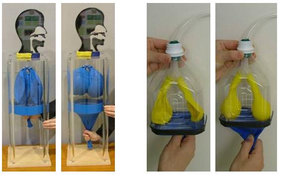
The lung models consist of a thoracic cavity, a rubber membrane, and two balloons. The rubber membrane covering the bottom of the thoracic cavity simulates the diaphragm. The two balloons inside the thoracic cavity simulate the lungs. The balloons are connected by a Y-shaped tube whose other end is attached to a whistle-type artificial larynx. Speech sounds are produced when the vocal tract model is connected to an artificial larynx.
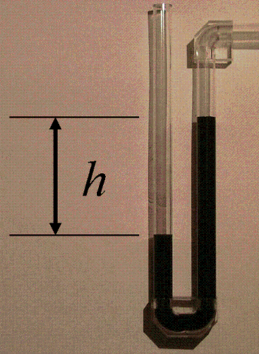
The first video clip shows a U-shaped tube with colored water attached to the thoracic cavity. When you look closely at the level of water in the U-shaped tube, the difference in height h reflects the pressure in the thoracic cavity. To set the vocal folds into vibration, the difference between the subglottal and supraglottal pressures, or the transglottal pressure, needs to be within the range of 5-10 cm H2O. In the video, h is within this range during phonation.
- Arai, T., “Lung model and head-shaped model with visible vocal tract as educational tools in acoustics,” Acoust. Sci. Tech., 27(2), 111-113, 2006.
- Arai, T., “Education system in acoustics of speech production using physical models of the human vocal tract,” Acoust. Sci. Tech., 28(3), 190-201, 2007.
- Stevens, K. N., Acoustic Phonetics, Cambridge, MA, MIT Press, 1998.
- Arai, T., “Virtual lung model for education in phonetics and speech science,” Acoust. Sci. Tech., 37(4), 173–174, 2016.